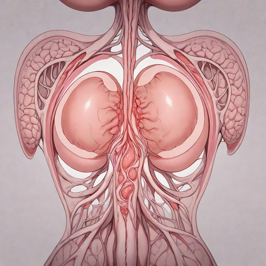
Fibroids are non‑cancerous growths that develop in or on the uterus and affect a large portion of women during their reproductive years. While many experience no symptoms, others face heavy menstrual bleeding, pelvic pressure, and fertility challenges. Understanding the condition, its causes, and the range of management options empowers you to make informed decisions with your healthcare team.
What Are Fibroids?
Fibroids, also called leiomyomas or myomas, are composed of smooth muscle cells and fibrous connective tissue. They can vary from microscopic seedlings to large masses that distort the uterus. Classification depends on location: intramural (within the uterine wall), submucosal (just under the inner lining), subserosal (on the outer surface), and pedunculated (attached by a stalk). Hormones, especially estrogen and progesterone, stimulate their growth, which explains why they often shrink after menopause.
Recognizing Common Symptoms
Not every fibroid produces noticeable signs. When symptoms appear they may include prolonged or heavy periods, anemia from blood loss, pelvic pain or pressure, frequent urination, constipation, backache, and pain during intercourse. In some cases fibroids interfere with implantation or increase the risk of miscarriage. Tracking menstrual patterns and any new discomfort in a journal helps your clinician assess severity and progression.
Diagnostic Pathways
A pelvic exam often provides the first clue. Imaging confirms size, number, and location. Transvaginal ultrasound is the most common first‑line tool; it offers clear pictures of the uterus and surrounding structures. For more detail, magnetic resonance imaging (MRI) maps fibroids in three dimensions and aids surgical planning. Hysteroscopy allows direct visualization of submucosal fibroids, while saline infusion sonography enhances detection of cavity‑distorting lesions.
Medical Management Options
When symptoms are mild, watchful waiting with regular monitoring may be appropriate. Pharmacologic therapies aim to control bleeding and shrink fibroids temporarily. Gonadotropin‑releasing hormone (GnRH) agonists induce a reversible menopause‑like state, reducing size but limited to short‑term use because of bone loss. Selective progesterone receptor modulators (SPRMs) such as ulipristal acetate can reduce volume and bleeding. Tranexamic acid and non‑steroidal anti‑inflammatory drugs (NSAIDs) lessen menstrual flow without affecting fibroid size. Hormonal intrauterine devices release levonorgestrel locally, decreasing heavy periods.
Minimally Invasive Procedures
Uterine artery embolization (UAE) blocks blood flow to fibroids, causing them to shrink. It preserves the uterus and typically requires an overnight stay. Magnetic resonance‑guided focused ultrasound surgery (MRgFUS) uses high‑intensity sound waves to destroy tissue without incisions. Radiofrequency ablation (RFA) delivers heat through a laparoscopic probe. These options suit women who wish to avoid hysterectomy and have fibroids amenable to targeted treatment.
Surgical Interventions
Myomectomy removes fibroids while leaving the uterus intact, preserving fertility. It can be performed hysteroscopically for submucosal lesions, laparoscopically for intramural or subserosal fibroids, or via an open abdominal incision for very large or numerous growths. Hysterectomy, the definitive cure, eliminates recurrence but ends childbearing potential. The choice depends on symptom burden, desire for future pregnancy, fibroid characteristics, and overall health.
Lifestyle and Complementary Strategies
While no diet or supplement cures fibroids, certain habits may ease symptoms. Maintaining a healthy weight reduces excess estrogen production from adipose tissue. A diet rich in fruits, vegetables, whole grains, and lean protein supports overall hormonal balance. Limiting red meat, alcohol, and high‑fat dairy may modestly lower risk. Regular aerobic exercise improves circulation and can lessen pelvic pressure. Stress management techniques such as yoga, meditation, or cognitive‑behavioral therapy help cope with chronic pain. Some women explore acupuncture or herbal remedies; discuss any supplement with your provider to avoid interactions.
Fertility Considerations and Pregnancy
Fibroids can affect conception and pregnancy outcomes, especially submucosal or large intramural types that distort the uterine cavity. Pre‑conception counseling often includes imaging to map fibroids and decide whether myomectomy improves chances. During pregnancy, fibroids may grow due to increased hormones, occasionally causing pain, preterm labor, or placental abruption. Close obstetric monitoring, pain management, and, rarely, surgical intervention during pregnancy are part of a tailored plan.
Long‑Term Follow‑Up and Monitoring
After any treatment, periodic ultrasound or MRI checks ensure fibroids have not regrown or new ones have not appeared. Symptom diaries, hemoglobin levels, and quality‑of‑life questionnaires guide ongoing care. Women approaching menopause often see natural regression, but hormone replacement therapy can stimulate residual growth; discuss risks and benefits with your clinician. Staying engaged with a multidisciplinary team — gynecologist, radiologist, fertility specialist, and primary care provider — optimizes long‑term health.
Shared Decision‑Making and Personalized Treatment Planning
Effective fibroid care begins with a structured conversation that aligns medical evidence with each woman’s values, reproductive goals, and lifestyle constraints. A shared decision‑making framework typically unfolds in three stages: information exchange, preference clarification, and joint selection. During information exchange the clinician presents a balanced overview of all viable options — watchful waiting, medical therapy, minimally invasive procedures, and surgery — using plain language, visual aids, and decision‑aid tools such as option grids or interactive calculators. Preference clarification invites the patient to rank outcomes that matter most: preserving fertility, avoiding major surgery, minimizing recovery time, controlling bleeding, or reducing long‑term hormone exposure. Joint selection then integrates these preferences with clinical factors such as fibroid size, number, location, and the presence of anemia or pressure symptoms. Documentation of the agreed plan in the electronic health record, including a follow‑up timeline and criteria for reassessment, ensures continuity when care transitions between primary care, gynecology, radiology, and fertility services. Edge cases — such as a patient who desires future pregnancy but has a large submucosal fibroid that distorts the cavity — may require a staged approach: hysteroscopic myomectomy first, followed by a trial of conception before considering more definitive therapy. Regular revisits, ideally at three‑month intervals after any intervention, allow the team to adjust the plan if symptoms recur, new fibroids appear, or life circumstances shift.
Emerging Pharmacologic Therapies and Clinical Trial Landscape
Beyond the established agents — GnRH agonists, selective progesterone receptor modulators, tranexamic acid, NSAIDs, and levonorgestrel intrauterine devices — several novel compounds are reshaping the therapeutic horizon. Oral GnRH antagonists such as relugolix and elagolix achieve rapid pituitary suppression without the flare effect of agonists, and they are now approved in combination with low‑dose estradiol and norethindrone acetate to mitigate bone loss and vasomotor symptoms. Early phase trials of selective estrogen receptor degraders (SERDs) and selective estrogen receptor modulators (SERMs) tailored for uterine tissue show promise in reducing fibroid volume while preserving endometrial integrity. Another frontier involves anti‑angiogenic agents — for example, bevacizumab‑based intra‑arterial delivery during uterine artery embolization — which aim to enhance ischemic shrinkage and lower recurrence rates. Women interested in investigational options should discuss eligibility criteria, required imaging, contraception mandates, and monitoring schedules with a research‑oriented gynecologist or a dedicated fibroid clinic. Practical considerations include insurance coverage for off‑label use, travel to trial sites, and the need for serial MRI assessments to capture volumetric response. Clinicians should maintain an up‑to‑date registry of recruiting studies (ClinicalTrials.gov, industry sponsor portals) and incorporate trial referral pathways into routine counseling, especially for patients who have exhausted standard therapies or who wish to preserve uterine function while avoiding surgery.
Racial, Ethnic, and Socioeconomic Disparities in Fibroid Care
Epidemiologic data consistently show that Black women develop fibroids at younger ages, experience larger tumor burdens, and report more severe symptoms than White, Hispanic, or Asian counterparts. These disparities arise from a complex interplay of genetic susceptibility, vitamin D deficiency, environmental exposures, chronic stress, and structural barriers to timely specialty care. Socioeconomic factors — limited insurance, transportation challenges, and health‑literacy gaps — further delay diagnosis and restrict access to fertility‑preserving procedures such as laparoscopic myomectomy or MRgFUS. Clinicians can mitigate inequities by implementing systematic screening questionnaires at primary‑care visits, offering telehealth triage for symptom assessment, and partnering with community health workers to navigate referral pathways. Culturally sensitive education materials, available in multiple languages and at appropriate literacy levels, improve understanding of treatment trade‑offs. Institutional policies that mandate equity audits — tracking time from symptom onset to imaging, to specialist consultation, and to definitive treatment — reveal hidden bottlenecks. For patients who face financial hardship, social‑work coordination can unlock medication assistance programs, charitable surgical funds, and transportation vouchers. Addressing these systemic issues not only improves individual outcomes but also enriches the evidence base by ensuring diverse participation in clinical trials and registries.
Pelvic Floor Physical Therapy and Sexual Health Rehabilitation
Fibroids frequently coexist with pelvic floor dysfunction, contributing to dyspareunia, urinary urgency, and chronic pelvic pain that persist even after fibroid reduction. A specialized pelvic floor physical therapist evaluates muscle tone, trigger points, coordination, and connective‑tissue mobility using internal and external manual techniques, biofeedback, and therapeutic exercise. Treatment plans often combine myofascial release, diaphragmatic breathing retraining, and progressive strengthening or down‑training depending on whether the patient presents with hypertonic or hypotonic patterns. For women reporting painful intercourse, therapists may introduce dilator therapy, graded exposure, and sensate focus exercises in collaboration with a sexual health counselor. Edge cases include patients with prior pelvic radiation, extensive adhesions from previous surgeries, or concurrent endometriosis; these require a multidisciplinary pain management approach that may incorporate nerve blocks, cognitive‑behavioral therapy, and pharmacologic neuromodulators. Integrating pelvic floor therapy early — ideally before any surgical intervention — can reduce postoperative pelvic pain, accelerate return to sexual activity, and improve overall quality‑of‑life scores. Insurance coverage varies; documenting medical necessity with validated outcome measures (e.g., Pelvic Floor Distress Inventory, Female Sexual Function Index) strengthens reimbursement claims.
Pre‑operative Optimization, Enhanced Recovery, and Post‑operative Surveillance
When surgery — whether myomectomy or hysterectomy — is chosen, a structured pre‑operative pathway reduces complications and shortens hospital stay. Optimization starts with anemia correction: intravenous iron or erythropoietin for hemoglobin below 10 g/dL, timed at least two weeks before the procedure. Nutritional screening identifies protein‑calorie deficits; a brief high‑protein supplement regimen improves wound healing. Smoking cessation at least four weeks prior lowers pulmonary and thromboembolic risk. For patients on GnRH agonists, a washout period of four to six weeks restores estrogen levels, decreasing vaginal atrophy and improving tissue pliability. Enhanced Recovery After Surgery (ERAS) protocols for gynecologic operations include preoperative carbohydrate loading, multimodal analgesia (scheduled acetaminophen, NSAIDs, gabapentinoids, and limited opioids), early ambulation within six hours, and early oral intake. Intraoperatively, meticulous hemostasis, uterine artery ligation when appropriate, and adhesion barriers (e.g., hyaluronic acid‑carboxymethylcellulose) lower postoperative adhesion formation. Post‑operative surveillance combines scheduled ultrasound at six weeks, three months, and twelve months with symptom diaries and hemoglobin checks. For myomectomy patients desiring pregnancy, a uterine cavity assessment — hysteroscopy or saline infusion sonography — before attempting conception confirms cavity integrity. Any new or enlarging fibroids trigger a repeat shared decision‑making session, ensuring the care plan evolves with the patient’s reproductive timeline.
Fertility Preservation Strategies Before Definitive Fibroid Treatment
Women who anticipate definitive fibroid therapy — such as hysterectomy, uterine artery embolization, or extensive myomectomy — and who wish to retain future biologic parenting options should be offered fertility preservation counseling early. The cornerstone is controlled ovarian stimulation with oocyte retrieval and vitrification, ideally completed before any gonadotropin‑suppressing medication or uterine artery occlusion. For patients with a partner, embryo cryopreservation after fertilization provides an alternative with known implantation potential. Timing is critical: a minimum of one menstrual cycle off GnRH agonists or antagonists allows follicular responsiveness to return. In cases where ovarian reserve is diminished (AMH <1 ng/mL, antral follicle count <5), a mild stimulation protocol or natural‑cycle retrieval may be employed to avoid excessive gonadotropin exposure. For women who cannot undergo stimulation due to time constraints or medical contraindications, ovarian tissue cryopreservation remains experimental but is available at select centers. Post‑preservation, a coordinated care plan outlines when to attempt conception — typically after uterine healing (three to six months post‑myomectomy, twelve months post‑UAE) — and includes pre‑implantation genetic testing if indicated. Counseling should also address the impact of fibroid location on implantation rates, the potential need for gestational carriers if uterine factor infertility persists, and the psychosocial aspects of delayed childbearing. Documentation of the fertility preservation discussion, consent forms, and a clear timeline in the medical record safeguards both the patient’s reproductive autonomy and the clinician’s medico‑legal responsibilities.
Advanced Imaging and Quantitative Assessment
Beyond standard transvaginal ultrasound and conventional MRI, several advanced imaging modalities add precision to fibroid mapping and treatment planning. Three‑dimensional (3D) ultrasound with volume rendering enables clinicians to measure total fibroid burden in cubic centimeters rather than relying on single‑plane diameters. Shear‑wave elastography provides a stiffness index that correlates with cellularity and predicts response to medical therapy; softer lesions tend to shrink more with GnRH antagonists. Contrast‑enhanced MRI using dynamic perfusion sequences quantifies vascularity, guiding decisions for uterine artery embolization versus focused ultrasound. Emerging AI‑driven volumetry platforms automatically segment multiple lesions on serial scans, reducing inter‑observer variability and allowing rapid calculation of percentage volume change after each intervention. In practice, a patient with numerous subserosal nodules may undergo baseline 3D ultrasound, followed by elastography at three months to gauge early pharmacologic response, and a contrast MRI before embolization to confirm feeder vessels. Edge cases include calcified fibroids that appear hypoechoic on ultrasound but hyperintense on T2‑weighted MRI; combining modalities prevents misclassification. Clinicians should document the imaging protocol, software version, and quality‑control metrics in the electronic record to ensure reproducibility across follow‑up visits.
Genetic Risk Profiling and Personalized Surveillance
While most fibroids arise sporadically, a growing body of evidence links specific somatic mutations and germline variants to tumor behavior. The MED12 exon 2 mutation appears in up to 70 percent of sporadic leiomyomas and correlates with hormone responsiveness. HMGA2 rearrangements and FH (fumarate hydratase) alterations identify hereditary leiomyomatosis and renal cell cancer syndrome, a rare but high‑risk condition warranting renal surveillance. Commercial polygenic risk scores now incorporate dozens of common SNPs associated with fibroid prevalence and severity, offering a quantitative estimate that can be shared during counseling. In a clinical workflow, a 28‑year‑old woman with a strong family history and a polygenic score in the 90th percentile might be offered annual MRI surveillance rather than biennial ultrasound, and earlier discussion of fertility preservation. Edge cases involve patients of mixed ancestry where reference panels are limited; clinicians should interpret scores cautiously and supplement with detailed pedigree analysis. When a pathogenic FH variant is identified, referral to a genetics counselor and coordination with nephrology become mandatory, illustrating how molecular data reshapes long‑term care pathways.
Navigating Insurance, Cost, and Access Barriers
Financial toxicity frequently determines which evidence‑based option a patient can pursue. Prior‑authorization requirements differ widely: GnRH antagonist combinations often need documented failure of first‑line agents, while uterine artery embolization may be classified as experimental by some payers despite guideline endorsement. Coding nuances matter — using CPT 58571 for laparoscopic myomectomy with morcellation versus 58573 for hysteroscopic resection influences reimbursement. A practical strategy is to create a pre‑visit checklist that captures diagnosis codes (ICD‑10 D25.9), planned procedure codes, and supporting documentation such as hemoglobin level, imaging reports, and failed conservative trials. Many institutions employ a dedicated financial navigator who can submit appeals, enroll patients in manufacturer assistance programs for relugolix‑estradiol‑norethindrone, and coordinate charitable funds for MRgFUS. Edge cases include uninsured patients who qualify for hospital charity care but face a waiting list for embolization; a bridge regimen of tranexamic acid plus an LNG‑IUD can control bleeding while the authorization process unfolds. Transparent cost estimators shared during shared decision‑making empower patients to weigh out‑of‑pocket expenses against clinical benefit.
Psychosocial Impact, Mental Health, and Support Resources
Chronic pelvic pain, unpredictable bleeding, and fertility uncertainty contribute to anxiety, depressive symptoms, and diminished sexual self‑esteem. Validated screening tools such as the PHQ‑9 and GAD‑7 should be administered at each fibroid‑focused visit, with scores triggering referral to a perinatal psychiatrist or a cognitive‑behavioral therapist experienced in chronic gynecologic conditions. Peer‑led support groups — both in‑person and virtual — have been shown to improve coping scores and reduce decisional conflict. A structured psychoeducational module covering realistic expectations for each treatment modality can be delivered in a single 60‑minute session before the shared decision‑making encounter. Edge cases include patients with prior trauma who experience pelvic exams as re‑traumatizing; offering a trauma‑informed approach with a chaperone, optional self‑insertion of the ultrasound probe, and the option to defer examination until trust is established mitigates avoidance behavior. Integrating a mental‑health liaison into the multidisciplinary fibroid clinic ensures seamless handoffs and documentation of psychosocial outcomes alongside clinical metrics.
Integrative Nutrition, Targeted Supplementation, and Lifestyle Prescription
Dietary patterns influence estrogen metabolism and inflammatory pathways that modulate fibroid growth. A Mediterranean‑style eating plan rich in cruciferous vegetables (indole‑3‑carbinol), green tea catechins (EGCG), and omega‑3 fatty acids has demonstrated modest reductions in leiomyoma volume in small randomized trials. Vitamin D deficiency (<20 ng/mL) is prevalent in women with symptomatic fibroids; repletion to 40‑60 ng/mL using 2000‑4000 IU daily cholecalciferol correlates with slower growth rates on serial MRI. Curcumin phytosome (500 mg twice daily) shows anti‑proliferative effects in vitro and is well tolerated, though drug‑interaction checks with anticoagulants are essential. Practical prescription: a 12‑week nutrition plan with weekly telehealth check‑ins, a food‑frequency questionnaire at baseline and week 12, and serum 25‑OH‑D monitoring at week 8. Edge cases involve patients on combined oral contraceptives where high‑dose vitamin D may alter hepatic metabolism; dose adjustment and liver function monitoring are advised. Exercise prescription should specify moderate‑intensity aerobic activity 150 minutes per week plus pelvic‑floor‑safe resistance training twice weekly, with progression guided by a physiotherapist familiar with fibroid‑related pelvic pressure.
Digital Health Tools, Telemonitoring, and Patient‑Reported Outcomes
Mobile applications and wearable sensors enable continuous symptom capture between clinic visits. A validated fibroid‑specific app can log daily menstrual flow using a pictorial blood loss assessment chart, record pain scores on a numeric rating scale, and sync with a Bluetooth‑enabled hemoglobinometer for home anemia screening. Data streams feed into a clinician dashboard that flags trends — such as a 30 percent rise in reported bleeding over two cycles — prompting early telehealth review. Remote video visits allow medication titration (e.g., adjusting relugolix dose) without travel burden, especially valuable for rural patients. Integration with the electronic health record via FHIR APIs ensures that patient‑reported outcome measures (PROMIS‑PF, EQ‑5D) become part of the longitudinal chart. Edge cases include limited digital literacy; providing a loaner tablet with pre‑installed app and a brief onboarding session improves adherence. Privacy safeguards — end‑to‑end encryption, HIPAA‑compliant cloud storage, and clear consent for data sharing — are non‑negotiable. Regular audit of app usage metrics informs quality‑improvement cycles and supports reimbursement for remote patient monitoring codes.
Transitioning Care Across Life Stages: Adolescence to Postmenopause
Fibroid presentation and management priorities shift dramatically from menarche through postmenopause. Adolescents may present with heavy menstrual bleeding and anemia; the first‑line approach emphasizes LNG‑IUD placement and tranexamic acid while preserving future fertility. Counseling must address body‑image concerns and school attendance, with a coordinated plan involving pediatric hematology for iron infusion if needed. In the reproductive years, fertility‑preserving myomectomy techniques (robotic‑assisted laparoscopic with ultrasonic dissection) are prioritized, and pre‑conception imaging timelines are aligned with assisted‑reproduction cycles. Perimenopausal women often experience rapid fibroid growth due to fluctuating estradiol; short‑course GnRH antagonist with add‑back therapy can bridge to menopause while minimizing bone loss. After menopause, asymptomatic fibroids typically regress, but hormone replacement therapy (especially estrogen‑only regimens) can reactivate growth; transdermal estradiol with micronized progesterone at the lowest effective dose is preferred, with annual MRI surveillance for the first three years. Edge cases include women who undergo surgical menopause before age 40; they require bone density monitoring, cardiovascular risk assessment, and a clear discussion about the trade‑offs of systemic versus local estrogen. A life‑stage roadmap documented in the chart guides each transition, ensuring that surveillance intensity, therapeutic goals, and patient education evolve in concert with physiologic changes.
Frequently Asked Questions
Can fibroids turn into cancer?
Fibroids are benign in the vast majority of cases. Leiomyosarcoma, a rare malignant tumor of smooth muscle, arises independently and is not considered a transformation of a typical fibroid. Routine imaging and pathology after removal provide reassurance.
Do all fibroids require treatment?
No. Asymptomatic fibroids discovered incidentally often need only observation. Treatment is guided by symptom severity, size, location, and reproductive goals.
Will a hysterectomy cure fibroids permanently?
Yes, removing the uterus eliminates the source of fibroids, so recurrence is impossible. However, it is a major surgery with permanent loss of fertility and potential hormonal changes if ovaries are also removed.
Can I shrink fibroids naturally without medication?
Evidence for natural shrinkage is limited. Weight loss, dietary changes, and stress reduction may modestly influence hormone levels and symptom burden, but they rarely cause significant size reduction on their own.
Is pregnancy possible after uterine artery embolization?
Pregnancy after UAE is possible, but studies show a higher rate of miscarriage, preterm delivery, and placental abnormalities compared with women who have not undergone the procedure. Discuss fertility preservation options with a specialist before choosing UAE.
Related posts:
 Why Black Women Face Greater Disparities With Fertility Treatment Option
 Understanding Blood Clots During Periods
 Your Complete Guide to Modern Fertility Treatment Options
 Pelvic Floor and Constipation
 The Hidden Financial Reality of Fertility Treatment
 The Importance of Fertility Education for Women’s Health
 New Tool Detects Melanoma Through Temperature
 ACL Injury Surgery: What You Need to Know