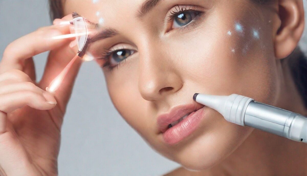
Understanding the Risks of Laser Skin Treatments
Cosmetic laser procedures have grown in popularity over the years as an effective way to improve skin concerns like wrinkles, sun damage, and unwanted hair. However, recent research indicates that certain laser treatments may increase the risk of developing skin cancer later in life.
How Do Lasers Damage Skin?
Laser treatments work by targeting water in the skin, which absorbs the laser energy and converts it into heat. This heat then destroys unwanted tissue. But in the process, DNA mutations can occur which may lead to cancer down the line.
Key Risk Factors to Consider
- Fairer skin types are more susceptible to laser damage and skin cancer in general
- People with a history of skin cancer in their family have a higher inherent risk
- The more laser treatments, the greater the cumulative risk
Protecting Yourself Going Forward
Rather than avoiding laser procedures altogether, it’s about being an informed patient. Have an open dialogue with your dermatologist about your unique risk profile. Start slow with less aggressive treatments. Apply broad-spectrum SPF 30+ sunscreen daily to protect your skin from further sun damage.
The Mechanics of Thermal Injury and DNA Integrity
While cosmetic lasers are generally classified as non-ionizing radiation—distinguishing them from the DNA-stripping power of X-rays or high-spectrum UV rays—the risk of cellular mutation arises through secondary thermal mechanisms. When a laser pulse strikes the skin, it aims for a specific chromophore, such as melanin, hemoglobin, or water. If the energy density (fluence) is too high or the pulse duration exceeds the ‘thermal relaxation time’ of the tissue, heat spills over into adjacent healthy cells. This phenomenon, known as collateral thermal damage, can induce a state of cellular stress that disrupts the normal replication cycle.
In this high-heat environment, the proteins responsible for DNA repair, such as p53, can be temporarily deactivated or overwhelmed. If a cell attempts to replicate while its DNA is compromised by heat-induced oxidative stress, it may incorporate errors into its genetic code. Over decades, these ‘silent’ mutations can accumulate. Practitioners often discuss the ‘bystander effect,’ where cells not directly hit by the laser beam receive chemical signals from damaged neighbors, leading to unexpected inflammatory responses. This suggests that the impact of a laser treatment isn’t just confined to the visible ‘spot’ but ripples through the surrounding architecture of the dermis, potentially altering the long-term genomic stability of the skin.
The Critical Danger of Masking Malignant Lesions
Perhaps the most immediate oncological concern regarding cosmetic lasers is not the creation of new cancer, but the accidental ‘masking’ of existing malignancies. Many patients seek laser therapy to clear up ‘age spots,’ ‘liver spots,’ or ‘sun-damaged patches.’ However, clinical studies have highlighted cases where a seemingly benign pigmented lesion was actually a melanoma in situ or an early-stage basal cell carcinoma. When a laser—particularly an Intense Pulsed Light (IPL) or a Q-switched laser—is used on these spots, it may successfully destroy the surface pigment without killing the underlying cancerous cells.
This creates a dangerous diagnostic vacuum. By removing the pigment, the laser takes away the primary visual cue that dermatologists use to identify skin cancer. The cancer may continue to grow vertically into deeper layers of the skin, hidden beneath a surface that now appears clear and rejuvenated. This ‘pseudo-clearance’ can delay a life-saving diagnosis by years. It is an absolute clinical necessity that any pigmented lesion intended for laser removal be examined via dermoscopy by a board-certified dermatologist first. If there is any structural asymmetry or irregular border, a biopsy must precede any cosmetic intervention to ensure that a malignancy isn’t being ‘shaved’ or ‘bleached’ rather than cured.
Ablative vs. Non-Ablative: Differing Mutagenic Profiles
The risk profile of a laser treatment is heavily dictated by whether it is ablative or non-ablative. Ablative lasers, such as CO2 or Erbium:YAG, work by vaporizing the entire top layer of the skin (the epidermis) and heating the underlying dermis. This process triggers a massive wound-healing response. While the complete removal of the epidermis can actually eliminate some pre-cancerous actinic keratoses—effectively ‘resetting’ the skin—the sheer intensity of the trauma requires the body to rapidly produce new cells. Any time there is rapid, forced cellular turnover in a high-inflammation environment, the statistical probability of a replication error increases.
Non-ablative lasers and fractional technologies (like Fraxel) create thousands of microscopic treatment zones (MTZs) while leaving the surrounding tissue intact. This ‘fractional’ approach is marketed as safer because it speeds up healing, but it creates a unique edge case: ‘field cancerization.’ If a patient already has significant sub-clinical sun damage, the laser might stimulate the proliferation of these damaged cells. Furthermore, the heat generated in non-ablative treatments stays trapped beneath the surface for longer periods, potentially leading to prolonged oxidative stress. Patients must weigh the ‘clean slate’ benefit of ablative procedures against the repeated, cumulative ‘insults’ of multiple non-ablative sessions, as the latter may contribute more to the total lifetime ‘mutational load’ of the skin.
The Role of Chronic Inflammation and the Microenvironment
Modern oncology increasingly views the ‘microenvironment’—the area surrounding a cell—as a key factor in cancer development. Cosmetic lasers, by design, induce controlled inflammation to stimulate collagen production. However, if treatments are performed too frequently or if the skin’s barrier is never allowed to fully recover, this can lead to a state of chronic low-grade inflammation. Chronic inflammation is a well-documented precursor to carcinogenesis; it involves the constant release of cytokines, growth factors, and reactive oxygen species (ROS) that can damage DNA and promote the survival of mutated cells.
Consider the case of ‘laser-induced Koebnerization.’ The Koebner phenomenon occurs when a skin condition (or potentially a malignancy) appears at the site of an injury. In patients predisposed to certain types of skin cancer, the physical trauma of the laser can act as a ‘trigger’ for a latent condition. Furthermore, the heat from the laser can alter the local immune landscape. The skin contains specialized immune cells called Langerhans cells that patrol for cancerous changes. Intense thermal energy can temporarily deplete or suppress these cells, creating a ‘window of vulnerability’ where the skin’s natural surveillance system is offline, allowing damaged cells to proliferate unchecked.
Identifying High-Risk Candidates Beyond the Fitzpatrick Scale
While fair skin (Fitzpatrick types I and II) is a primary risk factor, other physiological variables significantly influence how a person’s skin reacts to laser energy. Genetic predispositions, such as mutations in the MC1R gene (commonly associated with red hair and freckles), make the skin less efficient at repairing DNA damage following any thermal or UV insult. Even if a patient has a medium complexion, the presence of these genetic markers can heighten the risk of post-laser complications. Additionally, the use of photosensitizing medications—ranging from common antibiotics like tetracycline to certain blood pressure medications and even herbal supplements like St. John’s Wort—can cause the skin to absorb laser energy more aggressively than intended, leading to deep-tissue burns and increased mutagenic potential.
Immunosuppression is another critical edge case. Individuals on biologics for autoimmune conditions or those who have undergone organ transplants are at a much higher risk for skin cancers in general. For these patients, the inflammatory ‘trigger’ of a cosmetic laser may be more likely to catalyze a malignant transformation. It is also vital to consider the ‘UV history’ of the specific treatment area. Skin that has been chronically exposed to the sun, such as the décolletage or the tops of the hands, already has a high ‘mutational burden.’ Applying high-energy laser pulses to these areas requires extreme precision, as the cells are already on the brink of oncogenic transformation.
Post-Treatment Surveillance and the Atypical Healing Curve
Monitoring the skin after a laser procedure requires more than just checking for redness or peeling; it involves watching for ‘atypical’ healing patterns that could signal underlying issues. Normal healing follows a predictable trajectory: erythema (redness) and edema (swelling) should subside within a few days to two weeks, depending on the laser’s intensity. However, if a patient notices ‘islands’ of persistent redness, or if a specific spot becomes crusty, bleeds, or fails to heal after the rest of the face has recovered, this is a major red flag. These can be signs of a fast-growing squamous cell carcinoma triggered by the treatment’s inflammatory response.
Advanced surveillance protocols now recommend ‘total body photography’ and digital mole mapping for frequent laser users. By having a high-resolution baseline of the skin’s surface before starting a series of treatments, dermatologists can more easily spot new or changing lesions that might otherwise be dismissed as ‘just another laser scab.’ Patients should also be wary of ‘post-inflammatory hyperpigmentation’ (PIH) that does not resolve. While PIH is usually benign, it can sometimes mimic or hide ‘lentigo maligna,’ a slow-growing form of melanoma. A ‘wait and see’ approach is often dangerous; any pigment that returns in a different shape or color after a laser session should be treated with high clinical suspicion and undergo immediate professional evaluation.
The Regulatory Gap: Why the Operator Matters
The safety of a cosmetic laser is only as good as the person holding it. In many jurisdictions, the regulations governing who can operate a high-powered medical laser are surprisingly lax, leading to a rise in ‘med-spas’ where technicians with minimal medical training perform complex procedures. The risk of skin cancer is indirectly increased by improper settings; an inexperienced operator may use the wrong wavelength or an incorrect cooling duration, leading to ‘bulk heating’ rather than targeted treatment. This excessive heat is what most frequently leads to the DNA damage discussed earlier.
Furthermore, a non-medical operator is less likely to recognize the subtle signs of a pre-cancerous lesion. They may see a ‘brown spot’ and simply turn up the power to ‘blast it away,’ whereas a trained dermatologist would recognize the lesion as suspicious and pause for a biopsy. Equipment maintenance is another overlooked factor. Over time, laser mirrors can degrade and cooling systems can fail, leading to ‘hot spots’ in the laser beam that deliver uneven, dangerously high levels of energy to the skin. Choosing a facility that prioritizes medical oversight and rigorous device calibration is not just about getting better cosmetic results—it is a fundamental step in minimizing the long-term oncological risks of aesthetic medicine.
FAQ
What is the main risk from cosmetic lasers?
The main risk is developing skin cancer later in life due to DNA mutations from laser damage.
Who is most at risk from laser skin treatments?
Those with fair skin, family history of skin cancer, and who get multiple laser treatments over time have the highest risk levels.
Can I have any laser done safely?
Less aggressive laser treatments spaced out over time and paired with daily SPF use can minimize risk for most skin types.
What if I’ve already had many laser treatments?
Speak to your dermatologist about screening for signs of damage. Protecting your skin going forward is key to lower further risk.
Are other cosmetic skin procedures risky?
Laser resurfacing specifically has shown the most direct link to skin cancer risk compared to options like microneedling or chemical peels.
Related posts:
 Blow Drying Hair Without Damage: Expert Tips
 Cold Laser Therapy: A Non-Invasive Treatment Option
 Home Testing for Breast Cancer Risk
 Laser Hair Removal for Dark Skin: Debunking Myths and Sharing Success Stories
 Cancer Risk Factors for Endometriosis Patients
 Modern Medspa Wellness Trends
 New Tool Detects Melanoma Through Temperature
 HPV Test: The New Alternative to Pap Smears