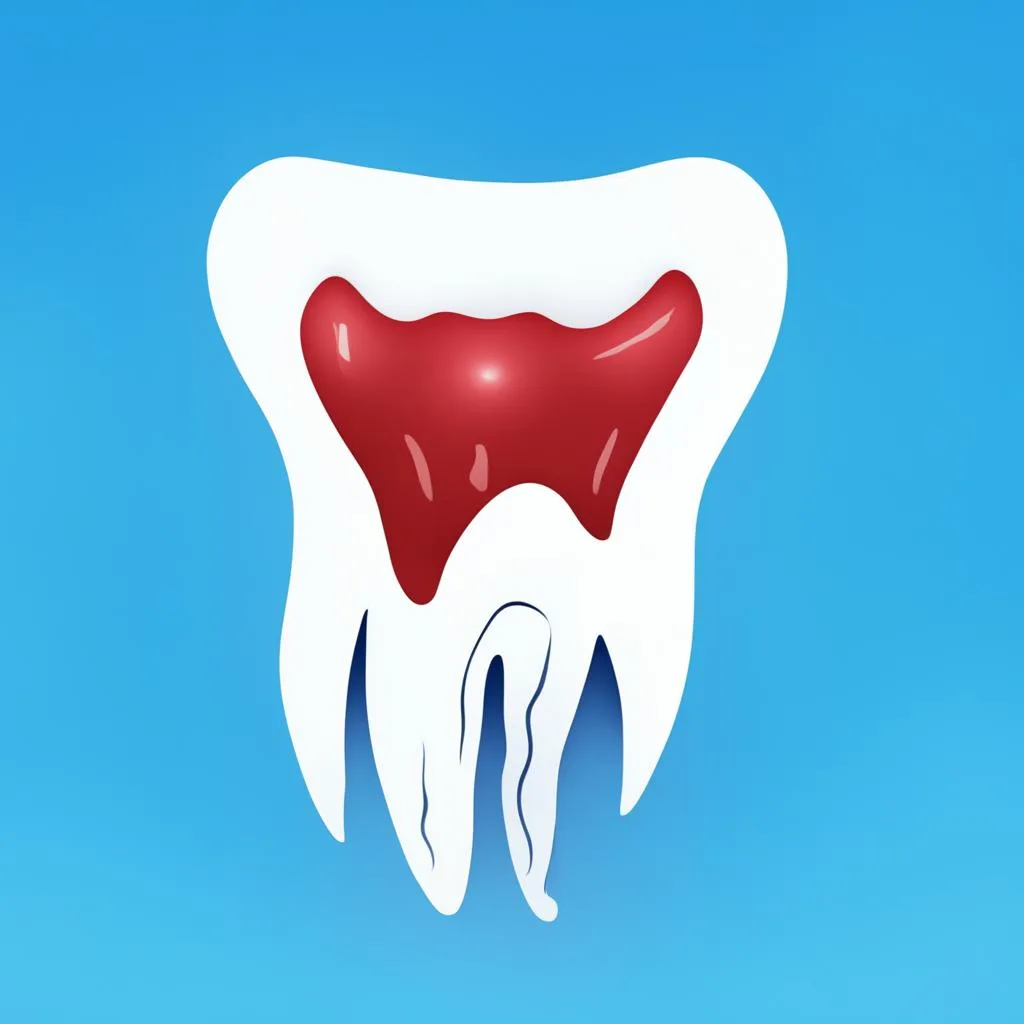
Root canals are often necessary when tooth decay has spread to the pulp – the innermost part of the tooth containing nerves and blood vessels. Instead of fear or anxiety over needing this procedure, it helps to approach it with compassion and understanding.
When is a Root Canal Needed?
There are a few common reasons why your dentist may recommend a root canal:
- Tooth decay has reached the pulp
- An injury or trauma has damaged the pulp
- The tooth is cracked and bacteria have reached the pulp
- Repeated dental procedures have irritated the pulp
Signs You May Need a Root Canal
Watch for these symptoms that indicate a root canal may be necessary:
- Severe tooth pain when eating or drinking something hot or cold
- Prolonged sensitivity to hot or cold foods and drinks
- Darkening of the tooth
- Pain and swelling
- Tenderness when chewing
- An abscess (pocket of pus) on the gums near the tooth
What Happens During a Root Canal?
During the procedure, the dentist removes diseased pulp tissue, cleans and shapes the inside of the tooth, then seals and fills the space. With modern techniques, root canals are often no more painful than having a filling placed.
Life After a Root Canal
With the diseased tissue removed and the tooth sealed, most patients find great relief from pain and discomfort. Some tenderness is normal during healing. Be gentle with the tooth initially, but you can resume normal brushing and flossing after a few days. With a restored tooth, you can enjoy chewing and smiling again.
Caring for Your Health Holistically
Good nutrition and oral hygiene help prevent tooth decay and the need for root canals. But when decay does happen or trauma occurs, modern dentistry can restore teeth to function and health. Approach the procedure with compassion towards yourself. Your dentist cares about preserving your wellbeing and your smile.
The Microscopic Battle: How Bacteria Navigate Dentinal Tubules
To understand why a root canal becomes necessary, one must look past the visible enamel and into the microscopic architecture of the tooth. Beneath the hard, protective enamel lies the dentin, a layer that is far more porous than it appears to the naked eye. Dentin is composed of thousands of microscopic channels known as dentinal tubules. These tubules radiate outward from the central pulp chamber to the exterior cementum or enamel border. When a cavity forms, it is not merely a hole; it is a breach in the tooth’s primary fortification. As decay progresses through the enamel, it reaches these tubules, which act as high-speed expressways for oral bacteria.
Once bacteria enter the dentinal tubules, they begin to migrate toward the pulp. The body’s initial response is to create “reparative dentin,” a denser layer intended to wall off the advancing infection. However, if the rate of bacterial invasion outpaces the tooth’s ability to produce this barrier, the microorganisms eventually reach the pulp chamber. This chamber houses the dental pulp, a delicate complex of nerves, blood vessels, and connective tissue. Because the pulp is encased in a rigid environment of dentin and enamel, it has no room to expand when it becomes inflamed. This lack of space is a primary driver of the intense pressure and pain associated with a tooth needing a root canal. The invasion of these tubules is often a slow process, meaning a patient might not feel discomfort until the bacteria are mere millimeters away from the nerve center.
In some edge cases, bacteria can reach the pulp even without a visible cavity. This occurs through microscopic cracks or through the gingival sulcus—the space between the tooth and the gum line. If a patient suffers from advanced periodontal disease, bacteria can travel down the side of the root and enter the pulp through the apical foramen (the opening at the tip of the root) or through accessory canals. This is often referred to as a “perio-endo” lesion, where the health of the gums and the health of the internal tooth are inextricably linked. Understanding this microscopic pathway highlights why early intervention with simple fillings is so critical before the bacterial colony secures a foothold in the tubule network.
Reversible vs. Irreversible Pulpitis: Identifying the Point of No Return
Dentists categorize pulp inflammation into two distinct stages: reversible and irreversible pulpitis. Distinguishing between the two is the deciding factor in whether a tooth requires a root canal or a more conservative treatment like a filling or a sedative dressing. Reversible pulpitis occurs when the pulp is irritated but still vital enough to recover if the source of irritation—such as a small cavity or a loose filling—is removed. In these cases, the pain is typically sharp but fleeting, lasting only a few seconds after a stimulus like cold water is introduced.
The transition to irreversible pulpitis marks the point of no return for the dental pulp. At this stage, the inflammation has caused significant tissue damage, and the blood flow within the pulp chamber is compromised. Because the pulp cannot heal itself once the bacterial load reaches a certain threshold, the tissue begins to undergo necrosis, or cellular death. Symptoms of irreversible pulpitis are often more severe and spontaneous; pain may linger for minutes or hours after the stimulus is gone, or it may wake a patient up in the middle of the night without any trigger at all. This spontaneous pain is a hallmark sign that the nerves are firing due to internal pressure and chemical mediators of inflammation rather than external contact.
Another clinical indicator used to diagnose irreversible pulpitis is the “lingering cold test.” During an examination, a dentist applies a specialized cold stimulus to the tooth. If the cold causes pain that persists for more than 30 seconds after the stimulus is removed, it suggests that the pulp’s vascular system is failing. In some instances, the tooth may even become sensitive to heat while being relieved by cold. This happens because heat causes the gases produced by decaying tissue to expand, increasing internal pressure, while cold causes them to contract, providing temporary relief. When a tooth reaches this stage, a root canal is the only way to remove the source of the infection and prevent the spread of bacteria into the surrounding jawbone.
The Silent Progression of Pulp Necrosis
One of the most dangerous misconceptions about root canals is that a lack of pain means the tooth is healthy. In reality, some of the most urgent cases involve pulp necrosis, where the nerve has already died. When the pulp tissue dies, the internal sensory mechanism is destroyed, and the acute pain of pulpitis may vanish. This often leads patients to believe that their tooth has “healed itself.” However, a necrotic tooth is essentially a hollow tube filled with dead tissue, which serves as a perfect breeding ground for anaerobic bacteria.
Without an active blood supply to deliver immune cells to the center of the tooth, the bacteria can multiply unchecked. Eventually, the infection migrates out of the apical foramen at the base of the root and into the periodontal ligament and alveolar bone. This is where a dental abscess forms. While the tooth itself may no longer feel sensitivity to temperature, it often becomes extremely tender to pressure or percussion (tapping). This is because the infection is now affecting the ligaments that hold the tooth in place. A patient might notice a small, pimple-like bump on the gums, known as a parulis or a sinus tract. This is the body’s way of creating a drainage port for the pus accumulating at the root tip.
Leaving a necrotic tooth untreated poses significant systemic risks. The chronic infection can lead to localized bone loss around the root, making the tooth mobile. In rare but serious cases, the infection can spread to the facial spaces, leading to cellulitis or even Ludwig’s Angina, a life-threatening condition that affects the airway. This is why dentists often recommend root canals for teeth that show signs of infection on an X-ray, even if the patient reports no current pain. The goal is to debride the dead tissue and seal the canal before an acute, painful flare-up occurs.
Impact and Insult: The Role of Physical Trauma and Thermal Shock
While decay is the leading cause of root canals, physical trauma is a frequent culprit, especially in younger patients. A sudden blow to the face—from a sports injury, a fall, or a car accident—can cause a tooth to undergo “concussion” or “luxation.” Even if the tooth does not break, the force of the impact can sever the delicate blood vessels entering the tooth at the root tip. Without this blood supply, the pulp becomes ischemic and eventually dies. This process can be immediate, or it can take years. A tooth that was bumped a decade ago may suddenly darken or develop an abscess, necessitating a root canal long after the original injury was forgotten.
Thermal shock is another less-discussed cause of pulp damage. The pulp is highly sensitive to extreme temperature changes. While rare, repeated exposure to high-heat dental procedures—such as the friction generated by a high-speed drill during the preparation of a large crown—can cause “iatrogenic” pulpitis. Modern dentists use water-cooling systems to mitigate this risk, but if a tooth has already undergone multiple large restorations, its ability to withstand further thermal stress is diminished. Each time a tooth is worked on, the pulp undergoes a degree of stress; over a lifetime, these “insults” to the tooth can have a cumulative effect, eventually pushing the pulp beyond its capacity to recover.
Furthermore, trauma can lead to a condition known as “calcific metamorphosis.” In response to an injury, the pulp may begin to rapidly deposit dentin along the walls of the canal in an attempt to protect itself. Over time, the canal can become almost entirely obliterated by calcified tissue. While this might sound like the tooth is strengthening itself, it often leads to the death of the remaining pulp tissue in a very narrow, confined space, making a subsequent root canal much more technically challenging for the dentist or endodontist. Early monitoring of traumatized teeth via regular X-rays and vitality testing is essential to catch these changes before they lead to infection.
Cracked Tooth Syndrome and Structural Compromise
Cracks in teeth are a common yet complex reason for root canal therapy. Unlike a visible chip in the enamel, “Cracked Tooth Syndrome” involves a fracture that often begins on the chewing surface and extends vertically toward the root. These cracks can be so fine that they are invisible to the naked eye and even difficult to detect on standard 2D X-rays. The pain associated with a cracked tooth is often unique: it typically occurs upon the release of biting pressure. This happens because the biting force opens the crack, and the release allows it to snap shut, irritating the pulp and the nerves within the dentin.
When a crack extends deep enough to reach the pulp chamber, it provides a direct gateway for bacteria. This is particularly problematic because the crack is a dynamic environment; every time you chew, the crack moves, pumping bacteria and debris deeper into the tooth. If the crack is limited to the crown of the tooth, a root canal and a full-coverage crown can often save the tooth. However, if the crack extends below the gum line or involves the floor of the pulp chamber, the tooth may be considered non-restorable. In these cases, the structural integrity is too compromised to support a root canal, and an extraction might be the only option.
Another structural issue involves “infractions,” which are craze lines in the enamel that don’t necessarily lead to the pulp. Distinguishing between a harmless craze line and a deep fracture requires diagnostic tools like transillumination, where a bright light is shone through the tooth to see if the light is blocked by a fracture line. Dentists may also use a “bite stick” to isolate which cusp is causing the pain. Addressing a cracked tooth early is vital; if caught while the inflammation is still reversible, a crown might suffice. Once the crack has contaminated the pulp, the root canal becomes the necessary intervention to stop the pain and save the tooth’s foundation.
The Importance of Advanced Diagnostic Modalities
Determining the need for a root canal is rarely based on a single symptom. Dentists use a battery of tests to confirm the health of the pulp. Beyond the visual exam and the cold test, Electric Pulp Testing (EPT) is a common tool. An EPT device delivers a tiny, controlled electric current to the tooth. If the patient feels a tingling sensation, it indicates the nerves are still functioning. A lack of response usually confirms pulp necrosis. However, EPT can sometimes yield false positives or negatives—for instance, if a tooth has a large metal filling that conducts the current to the gums, or if a multi-rooted tooth has one dead root and one live one.
Radiographic imaging is the cornerstone of endodontic diagnosis, but traditional 2D X-rays have limitations. They can show a dark shadow at the root tip (a periapical radiolucency), which indicates bone loss due to infection. However, because a 2D X-ray collapses a 3D object into a flat image, some infections can be hidden behind the root or the jawbone. This is where Cone Beam Computed Tomography (CBCT) has revolutionized the field. A CBCT scan provides a high-resolution 3D view of the tooth and surrounding bone. It allows the dentist to see the exact shape of the canals, identify hidden fractures, and detect infections that are not yet visible on traditional X-rays. This precision ensures that a root canal is only performed when truly necessary and increases the success rate of the procedure.
In addition to imaging, dentists use palpation and percussion tests. Palpation involves feeling the gum tissue near the root tip for tenderness or swelling, while percussion involves gently tapping on the tooth. Sensitivity to these tests suggests that the inflammation has spread beyond the tooth and into the surrounding periodontal ligament. By combining the results of temperature tests, EPT, 3D imaging, and physical exams, the dental team can create a definitive diagnosis. This comprehensive approach ensures that the source of the pain is accurately identified, especially in cases of “referred pain,” where a problem in an upper molar might feel like it is coming from the lower jaw.
Preventing Reinfection through Superior Coronal Sealing
The success of a root canal depends not only on the procedure itself but also on what happens afterward. A common cause for the “failure” of a root canal is not the internal cleaning, but rather “coronal leakage.” This occurs when the temporary or permanent restoration on top of the tooth fails to provide a hermetic seal. If saliva—which is loaded with bacteria—leaks under a filling or crown, it can travel down the root canal filling material (usually gutta-percha) and re-infect the area at the base of the root. This is why getting a permanent crown or high-quality filling placed promptly after a root canal is so critical.
Furthermore, because a tooth that has undergone a root canal no longer has a blood supply, it becomes more brittle over time. The dentin loses its moisture content, and the large access hole made to clean the canals further weakens the structural integrity of the tooth. Without a crown to “hoop” the tooth together and distribute biting forces evenly, a root-canaled tooth is at a much higher risk of fracturing. These fractures often occur vertically and can lead to the loss of the tooth even if the root canal itself was perfectly executed. A core buildup—a specialized filling that replaces the lost internal structure of the tooth—is often used to provide a solid foundation for the final crown.
In some complex cases, a dentist may use an endodontic post to help retain the core buildup. This is a small rod placed inside one of the cleaned root canals to provide extra support. However, modern bonding techniques have made posts less common than they once were. The transition from the root canal procedure to the final restoration is a delicate phase. Patients are often advised to avoid chewing on the treated side until the permanent crown is in place. By ensuring a tight, bacteria-proof seal at the top of the tooth and protecting the remaining structure with a crown, the tooth can function effectively for a lifetime, effectively ending the cycle of decay and infection that led to the need for the root canal in the first place.
Frequently Asked Questions
What are the signs I may need a root canal?
Severe tooth pain from hot or cold foods, prolonged temperature sensitivity, darkening of the tooth, swelling, tenderness, or an abscess can indicate pulp inflammation and the need for a root canal.
Is a root canal painful?
With modern techniques, most patients report root canal treatment as no more painful than getting a regular filling placed. Some tenderness during healing is common.
How long does a root canal take?
Most root canal treatments take 1-2 appointments of 60-90 minutes each. Simple cases may only require one visit. Follow-up restoration work like getting a crown adds more appointments.
Can any dentist do a root canal?
General dentists, endodontists (root canal specialists), and some pediatric dentists can all perform root canal treatment. Endodontists receive additional advanced education on root canal procedures.
How much does a root canal cost without insurance?
The cost for a root canal without insurance can range from $300-$2,000 depending on the tooth and complexity of treatment. Many dentists work with patients on payment plans as needed.
Related posts:
 Creating Your Own Crystal Crown
 Yes, Your Holistic Dentist Can Get Your Teeth Squeaky Clean
 Your Guide to Emergency Dental Care Solutions
 Consequences of Skipping Dental Checkups
 Products That Are Bad for Your Vaginal Health
 What Will a Dentist Do on a First Visit?
 Best Earplugs for Peaceful Sleep
 Sprained Tooth Guide